Cellular Imaging Systems
High-content imaging and analysis solutions, ranging from automated digital microscopy to high-throughput confocal imaging systems with water immersion objectives and proprietary spinning disk technology
An end-to-end solution for high-content imaging and analysis
Our systems for high-content imaging (HCI) and high-content analysis (HCA) provide flexible scalability making it easy to evolve your system alongside your research. They feature options and modules to address your specific research including objectives, filters, imaging modes, and environmental conditions. All our systems support a wide range of applications, increased throughput, and streamlined workflows.

Capture a diverse range of samples
Image live cells, stem cells, plants, tissue slices, whole organisms, and complex 3D matrices. The modularity and scalability of our platforms provide assurance that your system is a sound investment.

Analyze more data
Our industry-leading software solutions include powerful and elegant tools for imaging and analysis and offer flexibility and scalability.

Screen more assays
Our selection of imaging modes, application modules, analytics, and options for live-cell imaging combine to address hundreds of cell-based assays.
https://share.vidyard.com/watch/ZGN6gsqydWaZqDKXxGS4Ao
Explore a range of cell-based assays
Explore our high-content imaging portfolio
ImageXpress HCI system comparison
camera
objectives
autofocus
plates-supported
temperature-control
96-well-plate-scan-time-2-colors-1-site-well
maximum-illumination-channels
robotics-automation-compatible
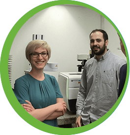
We like the user-friendly interface of the CRX [CellReporterXpress software with the ImageXpress Pico system] that makes it easy for non-specialists to use.
— Heather Martin, University of Leeds
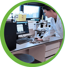
What we particularly like about our ImageXpress systems is the combination of flexibility and reliability. They are the workhorses of our group.
— Vardan Andriasyan, University of Zurich
The ImageXpress systems enable us to use our 3D organ-on-a-chip platform as a true high-throughput system. The ImageXpress Pico, ImageXpress Micro and ImageXpress Micro Confocal each provide valuable readouts, and all are a perfect match for the OrganoPlate®
— Jos Joore, MIMETAS
ImageXpress high-content imaging
(HCI) technology
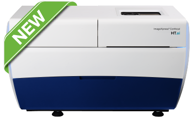
ImageXpress Confocal HT.ai system
Powerful multi-laser light sources, a deep tissue penetrating confocal disk module, water immersion objectives and modern machine learning analysis software
- Ideal for highly-complex cell-based and 3D assays
- Seven-channel high-intensity lasers generating brighter images with higher signal-to-background
- Spinning confocal disk technology for deeper tissue penetration, resulting in sharper images with improved resolution
- Water immersion objectives offering quadruple the signal at lower exposure times for greater sensitivity and image clarity without sacrificing speed
- Optional IN Carta software, leveraging intuitive, modern machine learning
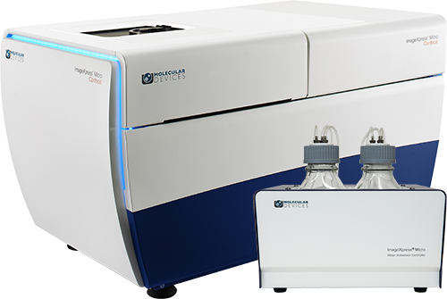
ImageXpress Micro Confocal system
High-content confocal imaging solution with proprietary spinning disk technology and water objective options
- Ideal for 3D organoid and spheroid imaging
- Proprietary AgileOptix™ spinning disk technology
- QuickID reduces image acquisition time and data storage requirements
- Water immersion objectives enhance resolution, sensitivity and throughput of complex
3D assays - Lasers and deep tissue confocal disk provide deeper penetration into 3D samples
- 3D volumetric analysis
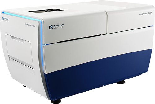
ImageXpress Micro 4 system
Configurable, high-throughput widefield imaging for fast biological processes
- Ideal for high-throughput screening, time-lapse imaging (from calcium assays to
multi-day subcellular assays) and intracellular yeast assays - Multiple imaging modes, including fluorescence, phase contrast, and brightfield
- >3 log dynamic range intensity detection
- QuickID reduces image acquisition time and data storage requirements
- Environmental control option for live cell assays and fast kinetic studies
- Field upgradeable to confocal
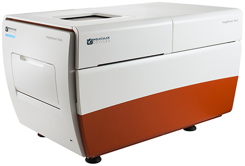
ImageXpress Nano
Fluorescence imaging, widefield platform for common biological assays
- Ideal for phagocytosis, mitotoxicity, autophagy, and cell differentiation
- Label-free brightfield imaging mode
- Up to five fluorescent filters can be installed, enabling multi-channel fluorescent and transmitted light imaging in one experiment
- High-speed autofocus
- Environmental control option for multi-day time lapse and live cell assays
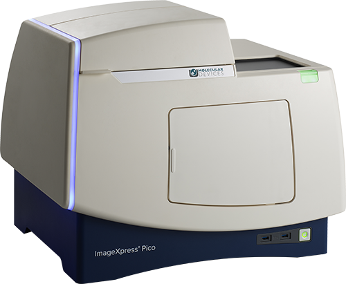
ImageXpress Pico system
Digital microscopy with automated brightfield, fluorescence, and real-time deconvolution imaging
- Ideal for cell counting, transfection efficiency, and cell health assays
- 25+ preconfigured application protocols
- 3D z-stack acquisition
- On-the-fly analysis
- Environmental control for live cell assays
- Access data from a browser— anytime, anywhere
- Optional Digital Confocal on-the-fly 2D deconvolution

Researchers gain new insights into immune response during pediatric respiratory infections using the ImageXpress Pico system
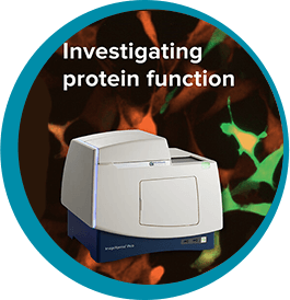
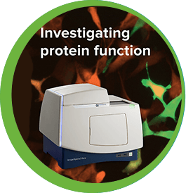
University of Leeds use ImageXpress Pico to investigate protein function

Forcyte Biotechnologies uses the ImageXpress Micro 4 system to help execute mechano-medicine discovery screens
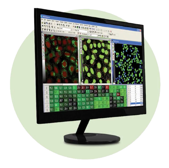
High-content image acquisition and analysis software
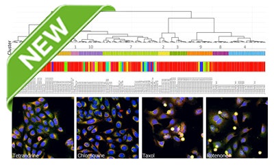
StratoMineR Advanced Cloud-Based Analytics
An intuitive and powerful platform for phenotypic profiling
Powerful and intuitive workflows allow users to import high-content imaging data directly into StratoMineR where it is used to generate rich, interactive visualizations using advanced data mining methods. When used with IN Carta Image Analysis Software, it provides robust, quantitative results from complex biological images and datasets utilizing advanced AI technology. Use all of your high-content data to discover, characterize, and analyze phenotypes.
MetaXpress software for ImageXpress high-content imaging systems
Multi-level analysis tools for a wide range of applications
- Meet high throughput requirements with a scalable, streamlined workflow
- Adapt your analysis tools to tackle your toughest problems, including 3D analysis
- Schedule automatic data transfer between third-party hardware sources and secure database
- Set up hundreds of routinely used HCS assays using MetaXpress software modules
- Export data to IN Carta software, leveraging intuitive, modern machine learning
Cell Image Gallery
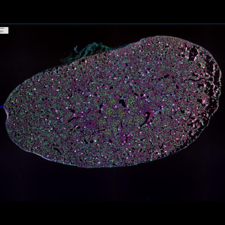
20 sites, stitched mouse kidney at 4X using the ImageXpress Nano system
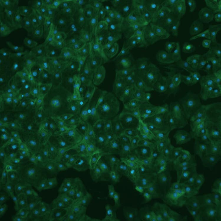
iCell cardiotoxicity using the ImageXpress Pico system
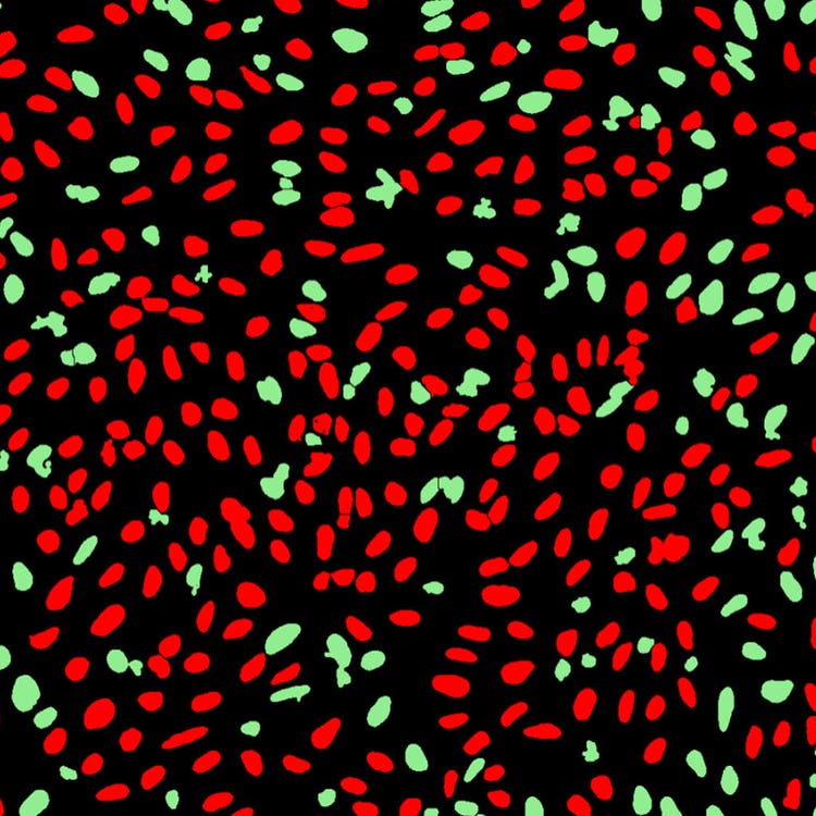
DNA damage mask in U2OS cells using the ImageXpress Nano system
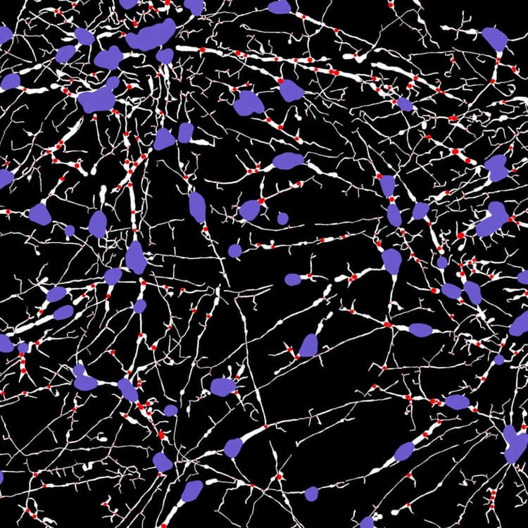
Stitched, neurite tracing mask at 40X using the ImageXpress Nano system
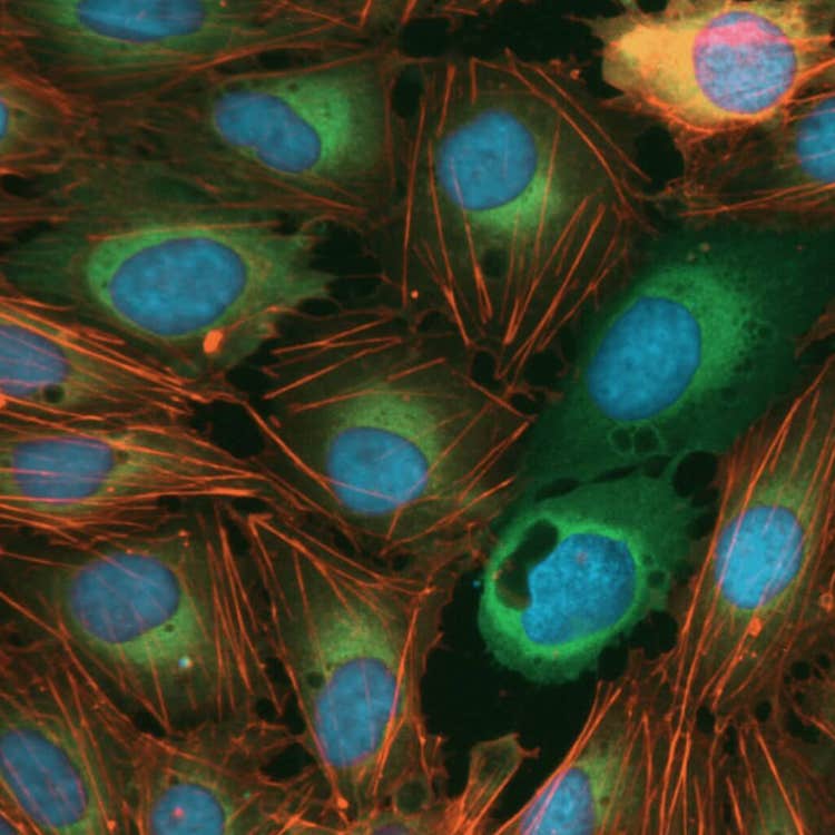
Cell morphology disruptors, acquired using the ImageXpress Nano system
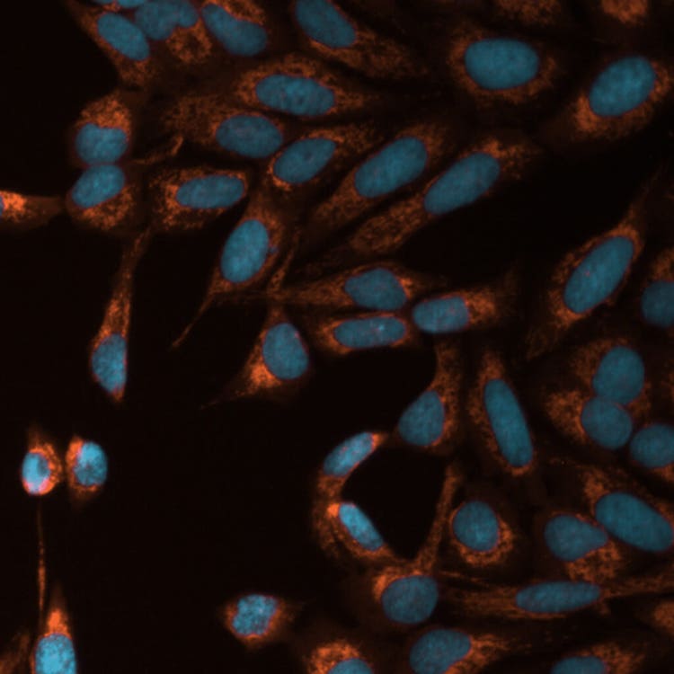
Mito toxicity with TRITC and DAPI using the ImageXpress Pico system
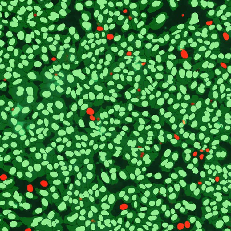
Live/dead control mask using the ImageXpress Pico system
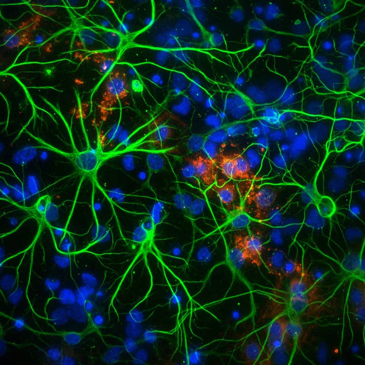
Neurons in 1536 well at 60X PA using the ImageXpress Micro Confocal system
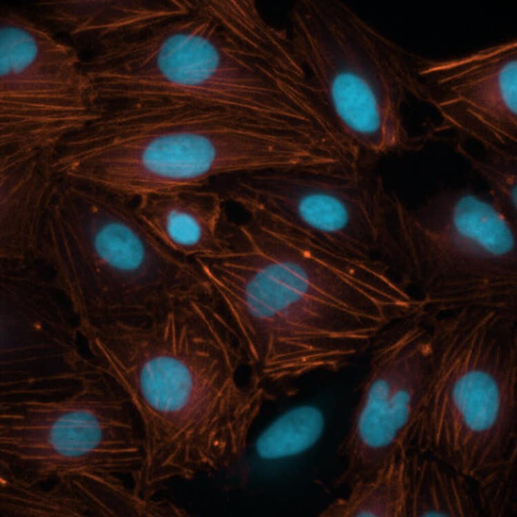
Nuclei using phalloidin stain at 60X using the ImageXpress Nano system
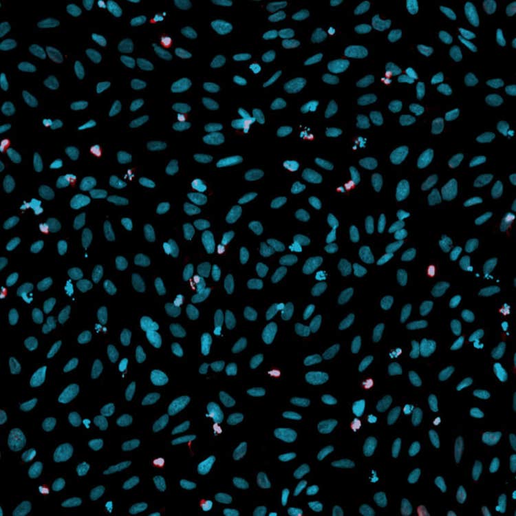
DNA damage overlay in U2OS cells using the ImageXpress Nano system
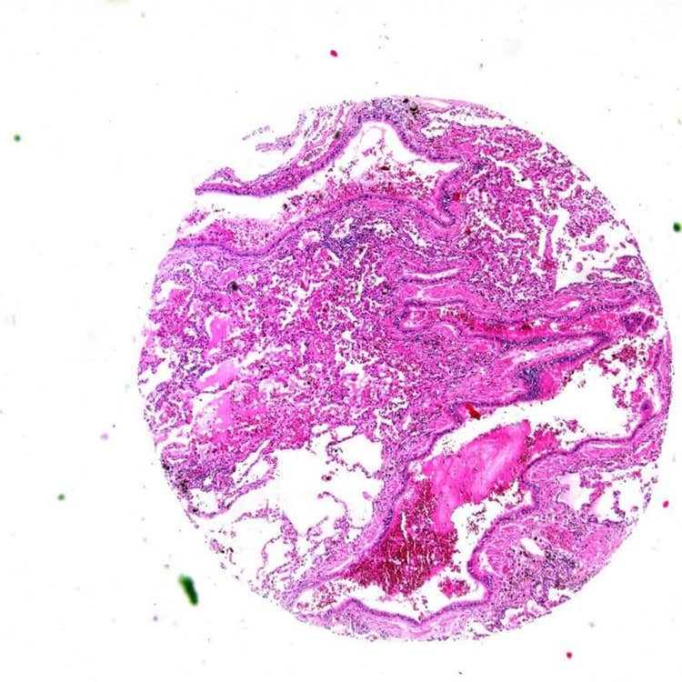
Array on a slide using the ImageXpress Pico system
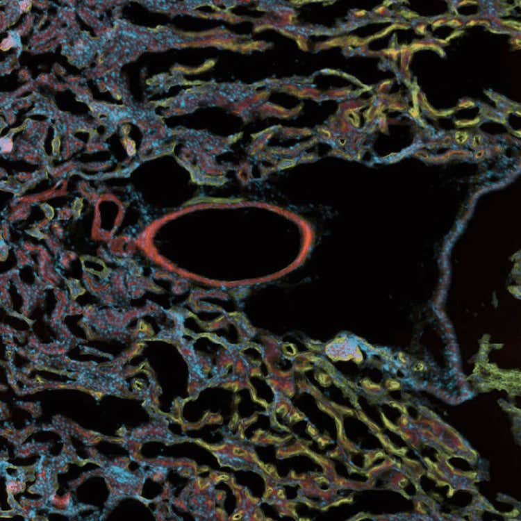
Fluorescent tissue.on a slide at 10X using the ImageXpress Pico system
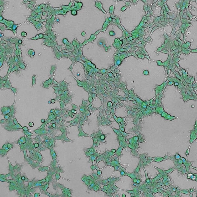
HEK293 live cell timelapse, 2 color using the ImageXpress Pico system
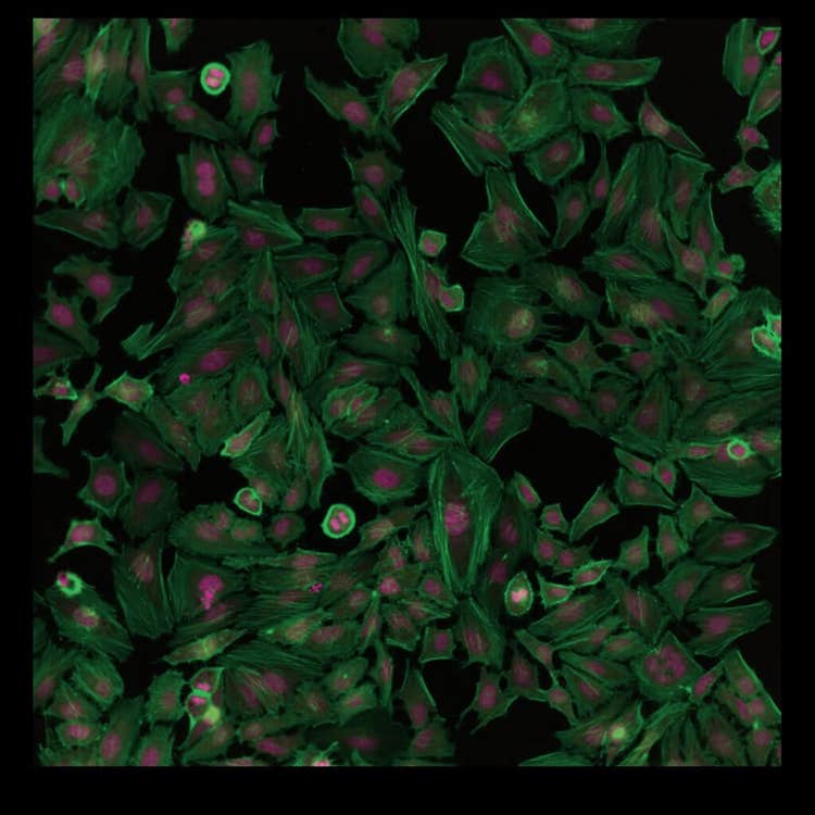
HeLa cell at 20X using the ImageXpress Pico system
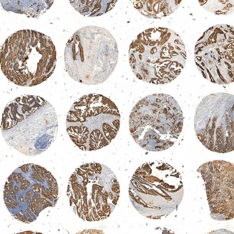
Array of tissue using the ImageXpress Pico system
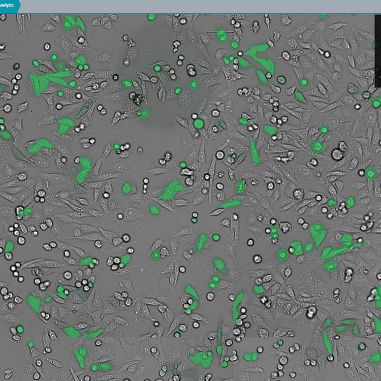
Transmitted light and GFP overlay using CellReporterXpress software on the ImageXpress Nano system
Enhance your imaging systems
With a variety of optional features and configurations for our high-content imaging systems, you can design a system to enhance your imaging capabilities. Our optional features enable access to a wider breadth of assays, additional capabilities for scaling basic and complex assays, and the ability to capture more physiologically-relevant cellular insights.

ENVIRONMENTAL
CONTROL

HIGH-THROUGHPUT 3D ANALYSIS

TRANSMITTED
LIGHT IMAGING

WIDE RANGE
OF OBJECTIVES
& FILTERS

ON-BOARD
ROBOTIC
FLUIDICS


Featured cell imaging applications
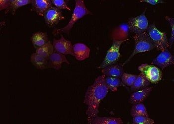
Cancer Research
Cancer involves changes which enable cells to grow and divide without respect to normal limits, to...
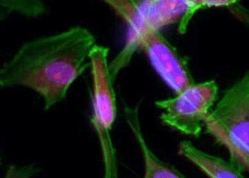
Cell Imaging
Researchers have several options in methods for imaging cells, from phase-contrast microscopy that...
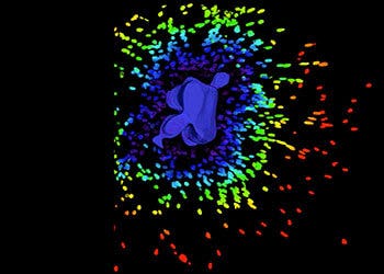
3D Cell Models
Development of more complex, biologically relevant, and predictive cell-based assays for compound..
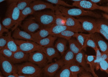
Live Cell Imaging
Live cell imaging is the study of cellular structure and function in living cells via microscopy....
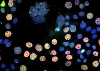
Cell Counting
Here we discuss the various methods and techniques used to assess proliferation, cytotoxicity, or...
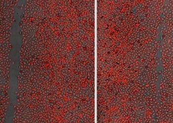
Cell Migration Assays
The movement or migration of cells is often measured in vitro to elucidate the mechanisms of...
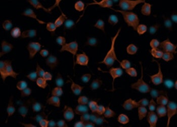
Neurite Outgrowth
Neurite outgrowth is assessed by the segmentation and quantification of neuronal processes. These...
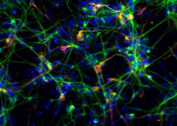
Stem Cell Research
Stem cells provide researchers with new opportunities to study targets and pathways that are more...
